Understanding Osteochondral Fractures: Causes, Symptoms, and Treatments
An osteochondral fracture is a condition where both the cartilage and the underlying bone within a joint are damaged. This type of injury most commonly occurs in weight-bearing joints such as the knee, but it can also affect other joints like the ankle, elbow, and shoulder. These fractures are usually the result of a traumatic event, such as a direct blow to the joint or a severe twisting motion. Understanding the intricacies of an osteochondral fracture is crucial for anyone facing this injury, as proper diagnosis and treatment can significantly impact long-term joint health and function.
What Is an Osteochondral Fracture?
An osteochondral fracture involves damage to two main components within a joint:
- Articular Cartilage: This smooth, elastic tissue covers the ends of bones in a joint, allowing for smooth movement and absorbing impact.
- Subchondral Bone: The bone layer just beneath the cartilage that provides structural support and stability to the joint.
When both of these structures are injured simultaneously, it results in what is known as an osteochondral fracture. These injuries can vary in severity, from small cracks in the cartilage to larger lesions where bone and cartilage detach from the joint surface.
Causes of Osteochondral Fractures
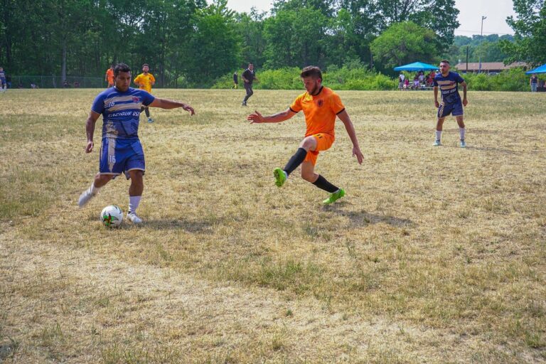
The primary cause of an osteochondral fracture is trauma. Sudden, forceful impacts, such as falls, sports injuries, or car accidents, are the most common sources. High-impact activities, especially those involving twisting or pivoting motions like basketball, soccer, or skiing, increase the risk of these fractures. Additionally, joint instability, prior injuries, or weakened bone and cartilage structures due to conditions such as osteoporosis or osteoarthritis can make individuals more susceptible.
Recognizing the Symptoms of Osteochondral Fractures
The symptoms of an osteochondral fracture can vary depending on the joint affected and the severity of the injury. However, common signs include:
- Pain: Sharp, localized pain at the site of the injury, especially during movement or weight-bearing activities.
- Swelling: Swelling around the affected joint, often accompanied by warmth or redness.
- Limited Range of Motion: Stiffness and difficulty moving the joint fully, particularly in severe fractures.
- Joint Locking or Catching: In cases where cartilage or bone fragments have become dislodged, the joint may lock or catch during movement.
- Instability: A feeling of instability or weakness in the joint, particularly in weight-bearing joints like the knee or ankle.
If left untreated, an osteochondral fracture can lead to chronic pain, joint degeneration, and a higher risk of developing osteoarthritis.
Diagnosis of Osteochondral Fractures
Accurate diagnosis of an osteochondral fracture is essential for determining the appropriate treatment plan. Physicians typically rely on a combination of physical examination and imaging tests to diagnose the condition. Common diagnostic tools include:
- X-rays: While X-rays can show bone fractures, they may not capture smaller cartilage damage, especially in early stages.
- Magnetic Resonance Imaging (MRI): MRIs are highly effective for visualizing both cartilage and bone injuries. They provide detailed images of the soft tissues in the joint, allowing for a comprehensive evaluation of the extent of the fracture.
- Computed Tomography (CT) Scans: CT scans are sometimes used to get a more precise view of the bone structures and any loose fragments in the joint.
- Arthroscopy: In some cases, a minimally invasive procedure called arthroscopy may be performed to directly visualize the joint and assess the damage.
Early detection of an osteochondral fracture is critical in preventing further joint deterioration and ensuring effective treatment.
Treatment Options for Osteochondral Fractures
The treatment of an osteochondral fracture depends on several factors, including the size of the fracture, its location within the joint, and the patient’s overall health and activity level. Treatment options range from conservative management to surgical intervention.
Conservative (Non-Surgical) Treatment
For minor osteochondral fractures, non-surgical options may be sufficient. These treatments typically include:
- Rest and Immobilization: Avoiding weight-bearing activities and immobilizing the joint with a brace or splint can help protect the joint and allow it to heal.
- Physical Therapy: Once the initial pain and swelling have subsided, physical therapy can help restore range of motion, strengthen surrounding muscles, and improve joint stability.
- Nonsteroidal Anti-Inflammatory Drugs (NSAIDs): Medications like ibuprofen or naproxen can help reduce pain and inflammation in the joint.
- Injections: In some cases, corticosteroid injections or hyaluronic acid may be used to reduce inflammation and improve joint function.
Conservative treatment is typically recommended for smaller fractures or for patients who may not be candidates for surgery due to health concerns.
Surgical Treatment
In more severe cases, or when conservative treatments fail to relieve symptoms, surgical intervention may be necessary. Common surgical procedures for osteochondral fractures include:
- Arthroscopic Debridement: This procedure involves using small instruments to remove loose cartilage or bone fragments from the joint. It can help alleviate symptoms but may not fully restore joint function.
- Osteochondral Autograft Transfer (OATS): In this technique, healthy cartilage and bone are harvested from another area of the patient’s joint and transplanted to the damaged area.
- Microfracture Surgery: Surgeons create tiny fractures in the subchondral bone to stimulate the formation of new cartilage. This procedure is often used for smaller lesions but may not be effective for larger or more complex fractures.
- Allograft Transplantation: For larger or more severe fractures, cartilage and bone from a donor may be transplanted into the damaged area.
- Autologous Chondrocyte Implantation (ACI): In this two-step procedure, healthy cartilage cells are harvested from the patient, cultured in a lab, and then implanted into the damaged area of the joint.
The choice of surgical procedure depends on the size and location of the fracture, as well as the patient’s overall health and activity level.
Rehabilitation and Recovery
Recovery from an osteochondral fracture can vary significantly depending on the severity of the injury and the treatment chosen. In cases of surgical intervention, recovery may take several months, and patients will need to follow a structured rehabilitation program.
Physical Therapy
Physical therapy is a critical component of recovery for most patients with osteochondral fractures. It helps restore joint function, improve flexibility, and strengthen the surrounding muscles. The intensity and duration of physical therapy will depend on the specific treatment and the patient’s progress.
Return to Activity
For athletes or individuals with physically demanding jobs, returning to full activity can take time. It is essential to follow medical advice closely and avoid rushing the process to prevent re-injury. In many cases, a gradual return to activity is recommended, with low-impact exercises introduced first, followed by more intense activities as the joint heals.
Long-Term Outlook and Complications
The long-term prognosis for patients with osteochondral fractures depends on several factors, including the size and location of the fracture, the patient’s age and overall health, and the promptness of treatment. While many patients recover fully and return to their regular activities, others may experience ongoing joint pain, stiffness, or instability.
One of the most significant long-term risks of an osteochondral fracture is the development of osteoarthritis. Damaged cartilage and bone can deteriorate over time, leading to chronic joint pain and reduced function. For this reason, early diagnosis and appropriate treatment are essential to minimize the risk of long-term complications.